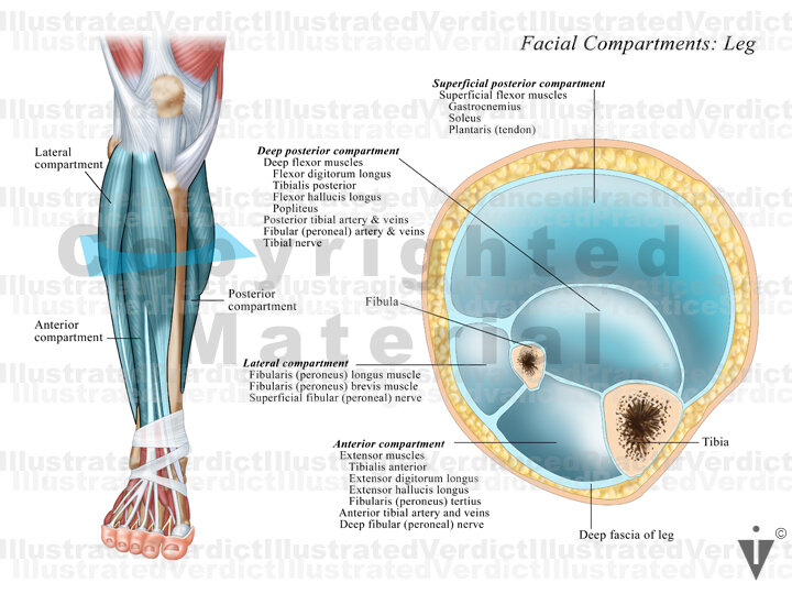
Necrotizing fasciitis is a clinical diagnosis, and imaging can reveal nonspecific or negative findings (particularly during the early course of disease). Mimics of necrotizing fasciitis include nonnecrotizing fasciitis (eosinophilic, paraneoplastic, inflammatory (lupus myofasciitis, Churg-Strauss, nodular, or proliferative), myositis, neoplasm, myonecrosis, inflammatory myopathy, and compartment syndrome. However, the lack of soft-tissue emphysema does not exclude the diagnosis. Dissecting gas along fascial planes in the absence of penetrating trauma (including iatrogenic) is essentially pathognomonic. Necrotizing fasciitis is a medical emergency with potential lethal outcome. Key imaging features are emphasized to enable accurate and efficient interpretation of variables that are essential in appropriate management. The purpose of this article is to review the imaging features of necrotizing fasciitis and its potential mimics. In elite athletes, MR imaging grading of muscle trauma plays an increasingly important role in recently developed comprehensive grading systems that are replacing the imprecise three-grade injury classification system currently used. In athletes, accurate grading of the severity and precise location of injury is necessary to guide rehabilitation planning to prevent reinjury and ensure adequate healing. The healing response after muscle trauma can result in regeneration, degeneration with fibrosis and fatty replacement, or disordered tissue proliferation as seen in myositis ossificans. Disorders related to the muscle's collagen framework include compartment syndrome, which is related to acute or episodic increases in pressure, and muscle herniation through anatomic defects in the overlying fascia. Direct impact to muscle results in laceration or contusion, often accompanied by intramuscular interstitial hemorrhage and hematoma. The risk of strain varies among muscles based on their fiber composition, size, length, and architecture, with pennate muscles being at highest risk. Strain is the most commonly encountered muscle injury and is characteristically located at the MTJ, where maximal stress accumulates during eccentric exercise. Concentric (shortening) contractions are more powerful, but it is eccentric (lengthening) contractions that produce the greatest muscle tension, leading to indirect injuries such as delayed-onset muscle soreness (DOMS) and muscle strain. This review illustrates the MR imaging appearance of a broad spectrum of acute, subacute, and chronic traumatic lesions of muscle, highlighting the pathophysiology, biomechanics, and anatomic considerations underlying these lesions. Magnetic resonance (MR) imaging is widely used for assessment of muscle injuries. The muscle and myotendinous junction (MTJ) are most commonly injured in the young adult, as a result of indirect mechanisms such as overuse or stretching, direct impact (penetrating or nonpenetrating), or dysfunction of the supporting connective tissues. Muscle is an important component of the muscle-tendon-bone unit, driving skeletal motion through contractions that alter the length of the muscle. It points out the affected compartments and allows the surgeon to selectively split the fascial spaces. MR imaging can help make the diagnosis of a manifest compartment syndrome in clinically ambiguous cases. Early follow-up showed changes in enhancement patterns late follow-up showed fibrosis and cystic and fatty degenerations of the affected compartments. T2-weighted spin-echo and magnetization transfer imaging showed bright areas, which enhanced after Gd-DTPA. Manifest compartment syndromes showed swollen compartments with loss of normal muscle architecture on T1-weighted spin-echo images. Early and late follow-up MR images were obtained.

In total, 15 patients (5 with an imminent compartment syndrome and 10 with manifest compartment syndrome) underwent MR imaging with a variety of pulse sequences including fat suppression, magnetization transfer imaging, and intravenous gadopentetate dimeglumine (Gd-DTPA) administration. The aim of this study was to evaluate the use of MR imaging for diagnosis and therapy management of compartment syndromes.


 0 kommentar(er)
